A letter in the New England Journal of Medicine October 2 by researchers affiliated with the Mayo Clinic reports on the Histopatholgy of Acute Lung Injury Associated with Vaping.
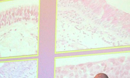
Despite the accumulating data on the clinical and imaging features of vaping-associated lung injury, its pathology is poorly understood. We reviewed lung biopsies from 17 patients (13 men; median age, 35 years [range, 19–67]), all of whom had a history of vaping (71% with marijuana or cannabis oils) and were clinically suspected to have vaping-associated lung injury. Presentation was acute or subacute in all cases, with bilateral pulmonary opacities; all but two patients presented in 2019. Eleven met the criteria for a “confirmed” diagnosis of vaping-related lung injury; the remaining six met the criteria for a “probable” designation. In all cases, histopathological findings showed patterns of acute lung injury, including acute fibrinous pneumonitis, diffuse alveolar damage, or organizing pneumonia, usually bronchiolocentric and accompanied by bronchiolitis (Figure 1; also see the Supplementary Appendix, available with the full text of this letter at NEJM.org). No histologic findings were specific, but foamy macrophages and pneumocyte vacuolization were seen in all cases and may be useful diagnostic clues in an appropriate clinical context. Pigmented macrophages were sometimes present but were never a dominant feature. Neutrophils were often prominent, but eosinophils were rare and granulomas were not seen. In two cases, bronchioloalveolar lavage fluid was available and contained abundant foamy macrophages. Despite treatment with glucocorticoids and maximum supportive care, two patients with diffuse alveolar damage died.
To date, few reports of vaping-associated lung injury have included histopathological findings. Our cases corroborate many of these descriptions and provide some preliminary insight into the pathogenesis of this injury. Much recent attention has been given to the possibility that vaping-associated lung injury may represent exogenous lipoid pneumonia. However, none of our cases showed histologic evidence of exogenous lipoid pneumonia and no radiologic evidence thereof has been found; this calls into question the diagnostic utility of identifying lipid-laden macrophages or performing oil red O staining on bronchioloalveolar lavage fluid as a marker of vaping-associated lung injury, as has been proposed. The significance of this observation remains unclear, particularly in patients with a known vaping history; until more data accumulate, our observations suggest that this finding should be interpreted with caution, as it may simply be a marker of exposure and not necessarily a marker of toxicity. Although it is difficult to discount the potential role of lipid, we believe that the histologic changes instead suggest that vaping-associated lung injury represents a form of airway-centered chemical pneumonitis from one or more inhaled toxic substances rather than exogenous lipoid pneumonia as such, but the agents responsible remain unknown.
Yasmeen M. Butt, M.D.
Maxwell L. Smith, M.D.
Henry D. Tazelaar, M.D.
Laszlo T. Vaszar, M.D.
Karen L. Swanson, D.O.
Mayo Clinic, Scottsdale, AZMatthew J. Cecchini, M.D., Ph.D.
Jennifer M. Boland, M.D.
Melanie C. Bois, M.D.
James H. Boyum, M.D.
Adam T. Froemming, M.D.
Mayo Clinic, Rochester, MNAndras Khoor, M.D., Ph.D.
Isabel Mira-Avendano, M.D.
Mayo Clinic, Jacksonville, FLAiyub Patel, M.D.
UnityPoint Health, Peoria, ILBrandon T. Larsen, M.D., Ph.D.
Mayo Clinic, Scottsdale, AZ
larsen.brandon@mayo.edu
Related news item: ASA’s Line on the Vaping Crisis